News and Press
ProScan Imaging Opens a New Multi-Modality Imaging Center in Liberty Township, Ohio
LIBERTY TOWNSHIP, OHIO – ProScan Imaging is excited to announce it is moving a hop, skip and a jump from its current location on Tylersville Road to a new larger facility offering expanded medical imaging services of MRI, CT and X-Ray.

ProScan Imaging Liberty will provide MRI, CT and X-ray at a lower cost than area hospitals, with patient-friendly enhancements including convenient front door parking, easy scheduling, evening appointment hours, and exceptional comfort and service.
ProScan Imaging’s mission is to help save patients’ lives through the use of advanced imaging technologies that support early and accurate diagnosis of disease. ProScan is the most experienced provider of outpatient MRI services in the Cincinnati region.
“We are excited to continue to raise the bar of patient care by offering a new best-in-class medical imaging center,” said Dr. Stephen Pomeranz, Founder and CEO of ProScan Imaging. “We have always been focused on providing local healthcare professionals and their patients with the best available medical imaging services and we are excited to continue expanding our efforts in Liberty Township”
The new facility in Liberty Township located on the roundabout of Cox Road, SR 129 and Veterans Blvd (across from Costco) is now open. The ProScan Tylersville MRI center closed on Monday, September 9.
The ProScan Family of Companies is headquartered in Cincinnati, Ohio and operates 31 independent medical imaging centers throughout the United States, including 11 locations in the Cincinnati/Northern Kentucky and Dayton areas. ProScan Imaging’s mission is to provide patients and providers with the highest-quality diagnostic medical imaging and radiology interpretations at a significantly lower cost compared to hospital imaging services. ProScan’s team of fellowship-trained, board-certified and sub-specialized radiologists are recognized as among the best and most experienced radiologists in the country.
About ProScan Imaging
For over three decades, ProScan Imaging has been a recognized leader in medical imaging and physician education in MRI. ProScan’s team of subspecialty trained radiologists are recognized as among the best and most experienced radiologists. ProScan Imaging is headquartered in Cincinnati, Ohio and provides teleradiology and on-site radiology services to physicians, imaging centers and hospitals in 45 US States and several US territories. The Company also owns and operates 30 independent imaging centers offering advanced imaging technology in multiple US States.
To learn more about ProScan Imaging, please visit: proscan.com.
For questions regarding this press release, please contact:
Rachel Seither Director, Physician Services & Corporate Marketing
Email: rseither@proscan.com Phone: 502-751-3668
New ProScan NCH Imaging Center Offers the First Contrast-Enhanced Mammography in Southwest Florida
An excerpt from The NCH Health Matters newsletter.
Naples, FL – One out of every eight women in the United States will develop breast cancer at some point in their lifetime. The first line of defense against this awful disease is early detection through quality imaging – performed routinely throughout every woman’s lifetime.
“In patients identified as high-risk, where the chances of breast cancer are increased, additional screening with MRI is recommended,” said Sergio Dromi, MD, ProScan NCH Director of Breast Imaging, board-certified and fellowship-trained in breast imaging.
ProScan Women’s Imaging at NCH provides the most comprehensive and cutting-edge imaging solutions, and just opened its newest facility on Vanderbilt Beach Road in North Naples. The facility offers all breast imaging services in one location including screening and diagnostic mammography (screening 3D/Tomosynthesis), MRI, ultrasound, contrast-enhanced mammography, and multimodality biopsies. It has been proven that 3D mammography improves the detection of breast cancer while decreasing the number of callbacks.
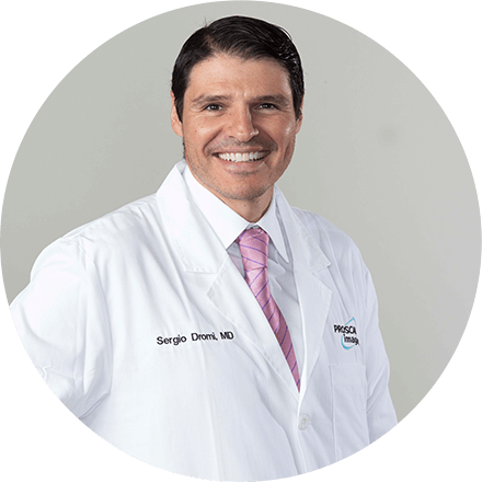
Sergio Dromi, MD, ProScan NCH Director of Breast Imaging, board-certified and fellowship-trained in breast imaging.
“ProScan Women’s Imaging at NCH is the first center in Southwest Florida to offer this imaging service,” explained Dr. Dromi.
Contrast-enhanced mammography works like a regular diagnostic mammogram with the addition of an injection of contrast to highlight areas of abnormal blood flow. Contrast will only be available for diagnostic mammograms. The facility also offers a dedicated 3T MRI for breast imaging that is faster and creates sharper images compared to low-field MRIs. The imaging center also offers dedicated ultrasound elastography, which helps distinguish between stiffness and soft tissues. “The majority of cancers demonstrate a stiffer behavior when compared to benign tissue, and elastography helps to determine whether or not a biopsy is needed,” said Dr. Dromi.
All imaging is performed by trained and certified technologists following the American College of Radiology and FDA guidelines. Dedicated breast imaging fellowship-trained radiologists interpret the images.
If previous breast imaging is immediately available, new screening exams will be read by a radiologist within 24 hours. If not, the patient’s prior imaging records are ordered, and the exam will be read within two weeks. “Prior exams are important in comparing the patient’s breast tissue year-over-year and help reduce unnecessary callbacks,” said Dr. Dromi.
“While the gold standard is still an MRI, we have launched the latest imaging tool for patients who cannot get an MRI; a contrast-enhanced mammography.”
– Sergio Dromi, MD, ProScan NCH Director of Breast Imaging
Guidelines state that patients with an average risk for breast cancer should start screening mammograms at the age of 40 – and should be repeated every year until life expectancy is no more than ten years.
“High-risk patients who have either a genetic predisposition, family history, or who test more than 20% on the Tyrer-Cuzick scale need to start the surveillance ten years before the first-degree relative was diagnosed with breast cancer – or at the age of 30. These patients should be screened every year with mammograms and MRI,” Dr. Dromi said.
The Tyrer-Cuzick scale provides a risk score that estimates the likelihood of a woman developing breast cancer in 10 years and throughout her lifetime.
“Patients with dense breast tissue have an indirectly increased risk for breast cancer due to the possibility of cancer being obscured by dense tissue. Interpreting these images is more complex, so additional surveillance with a more sensitive test, such as an MRI, is recommended,” Dr. Dromi said.
Screening mammograms can be self-referred. However, a physician or specialist referral is needed for diagnostic examinations.
Early detection of breast cancer is vital, because it can be treated easily with less aggressive surgeries and therapies. “A 3 millimeter cancer is much more easily excised and treated – with a much better long-term prognosis – than a 5 centimeter cancer,” Dr. Dromi added.
For more information or to schedule an appointment, call ProScan NCH at (239) 624-4443, option 3. The center is located at 2320 Vanderbilt Beach Road, Suite 1, in Naples.
ProScan Imaging Expands Open MRI and CT Services in Kentuckiana
Jeffersonville, IN – On December 18, 2023, ProScan Imaging, a renowned leader in advanced medical imaging, acquired OpenSided MRI of Jeffersonville, Indiana. The center, which is now owned and operated by ProScan Imaging, is located at 2027 Jeffersonville Commons Drive and features a state-of-the-art High-Field Fuji Oasis 1.2T Open High-Field MRI and Fuji Supria Plus CT.
This will be ProScan Imaging’s second location in the area. The company also offers MRI services at its outpatient imaging center in St. Matthews at 4044 Dutchmans Lane, Louisville, KY 40207.
“We are excited to expand our services for patients of Greater Louisville and Southern Indiana, providing cutting-edge technology at an affordable cost for both MRI and CT services.” said Karen Amaya, Chief Operating Officer of ProScan Imaging. “We are grateful for the overwhelming support the Louisville community has shown our St. Matthews center, and we’re thrilled for the opportunity to serve even more patients in the area.”
The ProScan Family of Companies is headquartered in Cincinnati, Ohio and operates 34 independent medical imaging centers throughout the United States. ProScan Imaging’s mission is to provide patients and providers with the highest-quality diagnostic medical imaging and radiology interpretations at a significantly lower cost compared to hospital imaging services.
ProScan’s team of fellowship-trained, board-certified, and sub-specialized radiologists is recognized as among the best and most experienced in the country.
“Being part of the Kentuckiana community has been an instrumental experience for our organization. We are excited to continue to raise the bar of imaging care by offering modern imaging modalities, including a High-Field Open MRI,” said Dr. Stephen Pomeranz, Founder and CEO of ProScan Imaging. “We have always been focused on providing local healthcare professionals and their patients with the best available medical imaging services, and we are excited to expand our efforts in Southern Indiana.”
The phone number and address of this location remain active. The phone number is (812) 282-0167 and the full address is 2027 Jeffersonville Commons Drive, Jeffersonville, Indiana 47130.
To learn more about ProScan Imaging Jeffersonville, please visit: https://proscan.com/
For questions regarding this press release, please contact:
Rachel Seither
Director, Physician Services & Corporate Marketing
Email: rseither@proscan.com
Phone: 502-751-3668.
CMS Lifts Coverage Limits on Amyloid PET
for Patients with Alzheimer’s Disease
As of today, CMS has lifted limitations and approved the local MACs to set up coverage determinations for amyloid PET/CT. This means that patients no longer need to be a part of a clinical trial under the prior CED (coverage with evidence development), and that multiple amyloid PETs will likely be covered in a patient’s lifetime.
This is great news, as we all know the importance of characterizing a patient’s neurodegenerative disorder with more accuracy to improve our care of this critical disease, especially with recent advancements in anti-amyloid therapy.
ProScan Imaging at NCH is already equipped with a new, top of the line PET scanner and staffed to meet the increased demands for amyloid PET that will come with this announcement.
Led by our Nuclear Medicine Director Michael Shriver, MD, along with his colleagues such as Dr. Jefferey Lieberman, our nuclear medicine physicians already have extensive experience in the interpretation of amyloid PET/CTs, having interpreted numerous studies over the past 5 years.
Please refer to the links below for more information on this exciting announcement. Feel free to reach out to Dr. Shriver via email (mshriver@proscan.com) if you have any questions about the nuclear medicine PET program at ProScan Imaging at NCH.
We look forward to assisting in the care of your patients.
CMS Decision Memo: https://www.cms.gov/medicare-coverage-database/view/ncacal-decision-memo.aspx?proposed=N&ncaid=308
ProScan Imaging Expands MRI and X-Ray Services
in the Greater Dayton Area
Vandalia, Ohio – On August 2, 2023, ProScan Imaging, a renowned leader in advanced medical imaging, relocated its imaging center in Troy, Ohio to a new site in Vandalia, Ohio. The new center, located at 810 Falls Creek Drive, Suite A in Vandalia, features a state-of-the-art high-field Fujifilm Echelon Oval 1.5T MRI and digital X-ray.
“We are excited to expand our services to patients of North Dayton providing cutting-edge technology at an affordable cost for not only MRI services, but X-Ray as well.” said Dr. David Griffith, Medical Director of ProScan Imaging Vandalia. “The support that the Troy/Dayton community has shown us over the past 17 years has been overwhelming and this has inspired us to expand our services into Vandalia.”
The ProScan Family of Companies is headquartered in Cincinnati, Ohio and operates 33 independent medical imaging centers throughout the United States, including 12 locations in the Dayton/Cincinnati/Northern Kentucky area. ProScan Imaging’s mission is to provide patients and providers with the highest-quality diagnostic medical imaging and radiology interpretations at a significantly lower cost compared to hospital imaging services. ProScan’s team of fellowship-trained, board-certified and sub-specialized radiologists are recognized as among the best and most experienced radiologists in the country.
The addition of the new Vandalia imaging center is significant for the local community as patients now have an independent, lower-cost and easily accessible imaging option for MRI and X-Ray services in the Greater Dayton region.
“Being part of the fabric of the Troy and Greater Dayton community has been an instrumental experience for our organization. We are excited to continue to raise the bar of imaging care by offering modern imaging modalities,” said Dr. Stephen Pomeranz, Founder and CEO of ProScan Imaging. “We have always been focused on providing local healthcare professionals and their patients with the best available medical imaging services and we are excited to expand our efforts in Vandalia.”
The new address of the ProScan Imaging Troy center is now 810 Falls Creek Dr, Suite A, Vandalia, Ohio 45377. The old address of 45 S. Stanfield, Suite 101, Troy, Ohio is no longer in service. However, the phone number remains active at 937-440-6781.
For a price quote or to schedule an MRI or X-Ray appointment at the new Vandalia location, visit our website at https://proscan.com/locations/vandalia/ or patients can call 937-440-6781.
![]()
About ProScan Imaging
For over three decades, ProScan Imaging has been a recognized leader in medical imaging and physician education in MRI. ProScan’s team of subspecialty trained radiologists are recognized as among the best and most experienced radiologists. ProScan Imaging is headquartered in Cincinnati, Ohio and provides teleradiology and on-site radiology services to physicians, imaging centers and hospitals in 45 US States and several US territories. The Company also owns and operates 33 independent imaging centers offering advanced imaging technology in multiple US States.
For questions regarding this press release, please contact:
Rachel Seither
Director, Physician Services & Corporate Marketing
Email: rseither@proscan.com
Cleerly Partners with ProScan Imaging to Transform the Standard of Care for Heart Disease Prevention
ProScan’s imaging centers in two major regions will now leverage Cleerly’s AI-enabled approach to develop personalized heart care diagnoses and treatment plans
NEW YORK – May 31, 2023 – Cleerly, the company creating a new standard of care for heart disease, announced today a partnership with ProScan Imaging to offer more personalized approaches to heart health, cardiovascular analyses, and treatment plans. ProScan Imaging is recognized as being at the forefront of using new imaging technology to deliver the best possible care to its patients. The company plans to use Cleerly’s AI-enabled platform to evaluate coronary computed tomography angiography (CCTA) imaging, allowing radiologists and cardiologists to identify, characterize and qualify atherosclerosis (plaque) buildup in the walls of heart arteries more easily. The personalized reporting provided through this platform allows providers to deliver precision treatment across the continuum of heart disease severity, improving clinical outcomes while drastically reducing costs. “We are thrilled to be partnering with ProScan Imaging on their mission to deliver the best possible diagnostic care to their patients,” said James K. Min, MD, FACC, FESC, MSCCT, founder and CEO of Cleerly. “This partnership will allow us to improve heart health through a personalized approach to evaluating patients’ heart disease risk and continue to support Cleerly’s mission to create a world without heart attacks.”
Cleerly’s comprehensive coronary analysis provides actionable insights for physicians that go beyond traditional CCTA scans with detailed vessel-by-vessel evaluation of stenoses and atherosclerosis. Its heart disease technologies serve as an all-in-one comprehensive, coronary artery disease (CAD) evaluation solution. For every artery and its branches, Cleerly’s technologies accurately and precisely quantify atherosclerosis, vascular morphology, and assess vascular profiling. Through its four-tiered, atherosclerosis plaque-burden staging system, Cleerly describes patients based on total plaque volume or percent atheroma volume with stages that range from normal, mild, moderate, to severe plaque. Additionally, Cleerly evaluates changes in plaque burden through longitudinal disease tracking of changes in CAD over time.
These diagnostic tools and reporting solutions are tailored to all stakeholders in the heart care pathway – be they radiologists, cardiologists, primary care physicians, or patients and their caregivers. ProScan Imaging is one of the largest outpatient imaging networks in the country, with 33 centers in five regions, including Ohio, Indiana, Kentucky, New York and Florida. Upon launch, Cleerly services will be available in ProScan’s largest markets: Cincinnati, OH and Naples, FL imaging centers. This partnership will provide ProScan Imaging access to Cleerly’s care pathway to care for patients across the entire continuum of disease presentation – from early diagnosis of heart disease to ensuring that ongoing treatments are effective for those who have elevated risk of symptoms.
“This new partnership with Cleerly supports our mission to provide the highest-quality diagnostic care to patients,” said Stephen J. Pomeranz, MD, Medical Director and CEO of ProScan Imaging. “We are passionate about empowering patients and providers to make proactive and well-informed medical decisions, and the Cleerly analysis provides the insight needed to make better choices for heart health.” Cleerly’s services are now available at several ProScan Imaging centers and those interested can visit https://proscan.com/ to learn more and schedule an appointment.
About Cleerly
Cleerly is the company creating a new standard of care for heart disease. Through its FDA-cleared solutions driven by artificial intelligence, Cleerly helps enable comprehensive phenotyping of coronary artery disease, as determined from advanced non-invasive CT imaging. Led by a world-class clinical and technical team, Cleerly enhances health literacy for each and every stakeholder in the coronary care pathway. For more information, please visit: https://www.cleerlyhealth.com.
Cleerly Press Contact: press@cleerlyhealth.com
About ProScan Imaging
For over three decades, ProScan Imaging has been a recognized leader in medical imaging and physician education in radiology. ProScan’s team of subspecialty-trained radiologists are widely- recognized as among the best and most experienced radiologists. ProScan Imaging is headquartered in Cincinnati, Ohio and provides teleradiology and on-site radiology services to physicians, imaging centers and hospitals in 45 US States and several US territories. The Company also owns and operates 33 independent imaging centers offering advanced imaging technology in multiple US States.
To learn more about ProScan Imaging, please visit proscan.com/ct-scans/